|
 |
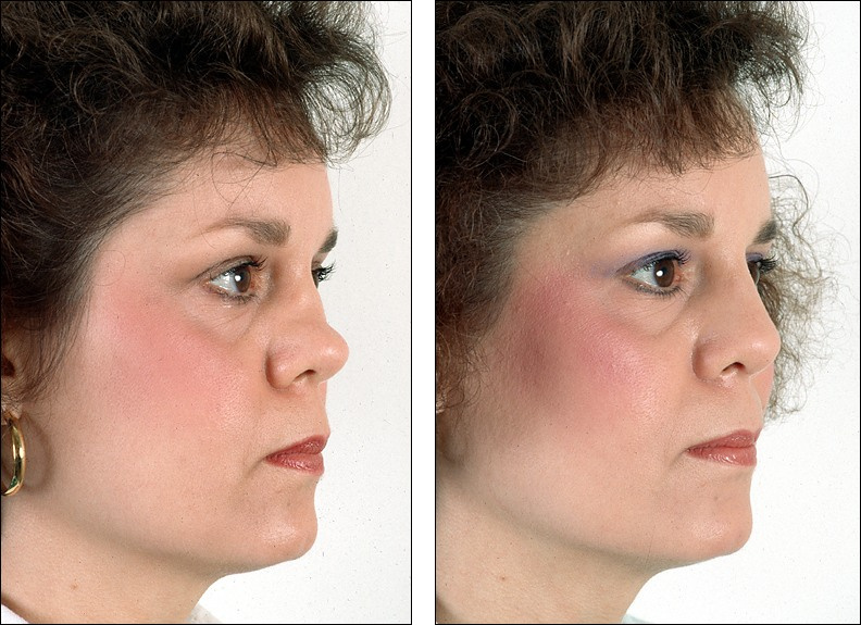
In the before picture, the cartilages that form the tip of her nose are fully
present. Above the tip cartilages is the divot resulting from loss of some
tissue in that area. Above the divot, her nasal bones are in their normal
position.
If you know where the nasal bones normally reside, you can see their outline
in the before picture above. If you'd like to find out more about the
location of the nasal bones and how they are addressed during surgery, check out
that chapter
of the rhinoplasty surgery tutorial (the surgery tutorials contain explicit photographs taken during surgery).
|
|
next view of this patient
|
| All views of this rhinoplasty patient: |
 Go here to learn how to send your photos to Dr. Denenberg, Go here to learn how to send your photos to Dr. Denenberg,
 or to arrange a personal consultation. or to arrange a personal consultation.
Next: an example of the solid advice Dr. Denenberg gives patients on RealSelf.com.
Get that advice for your own situation by emailing your photos to Dr. Denenberg.
|
Questioner:
My plastic surgeon doesn't use drain tubes after Neck Lift/Facelift. Is that okay?
(Questioner submitted photos)
Dr. Denenberg's answer: Drains after facelift
Deb,
This is a question that cosmetic surgeons discuss regularly and you will find many different opinions. Most feel drains do help minimize seromas (fluid collections under the skin) and improve the recovery process. Some surgeons use fibrin sealants which also work well. Others just depend on dressings and pressure. In general, however, if your surgeon is experienced with facelifts and getting excellent results, then whether or not they use drains should not be an issue.
Best of luck with your procedure-
Link to this question on RealSelf.com
|
|