wasperformed FacialSurgery.com
Steven M. Denenberg, M.D.
Dr. Denenberg's articles on Medium.com.

Rhinoplasty tutorial >> Hump removal >> page 11
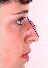
Image size: small
show larger
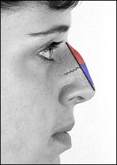
Remember this illustration?  Having removed the cartilaginous hump (shaded blue above right), our next task is to remove the portion of the hump that is made out of bone, the part colored pink in the diagram.  Since the nasal bones are part of the skull, we now need to look at a skull in order to understand how we work on those bones.

Click on any image in this tutorial to see a greatly-enlarged version
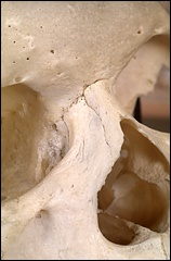
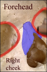
This photograph is a close-up of the part of the skull that we are dealing with in rhinoplasty.  The diagram above right will help to orient you.  The photograph was taken from the same perspective as the pictures of noses we've been studying: photographer standing to the skull's right.  The eye sockets are outlined in red, and the bones in the area of the nose are colored blue.


Clear all red checks in the Rhinoplasty Tutorial


All surgery depicted in this essay, except where noted, was performed by Dr. Denenberg