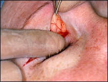
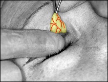
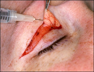
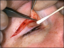
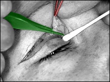
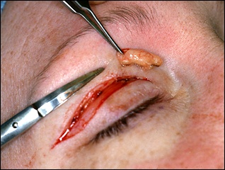
 The fat is now injected
with the anesthetic solution before excising it. When we injected the eye
before beginning surgery, we injected just underneath the skin, which
anesthetized the skin and the muscle that hugs the skin. However, this fat
was protected underneath the orbital septum membrane, and the anesthetic
solution didn't diffuse down to numb it. So we inject it now.
The patient is
snoozing, and she doesn't feel the placement of the teeny needle into the
fat. She would, however, feel the fat excision if we forgot to numb it
here.
|
