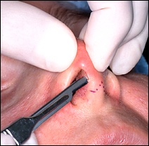
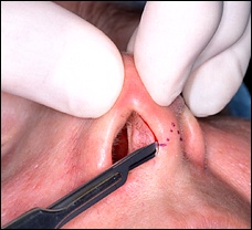
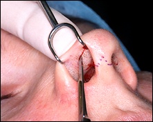
 Now I make the lateral
part of the marginal incision. The incision is called "marginal"
because it follows the margin of the nasal cartilage that gives the tip of the
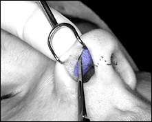
nose its shape. The image above right shows the position of that cartilage
in blue, and I'm trying to hug its lower edge with the scalpel. You'll see the entire cartilage
as soon as we finish the incisions.
In medical lingo,
"lateral" means "away from the middle," so this is
the lateral part of the incision. The "medial" part of the
incision, the part "closer to the middle," is the incision that
hugged the side of the columella, pictured at the top of this page.
|
